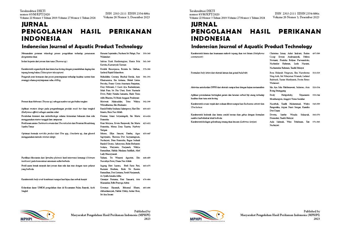
Isolasi heparin dari jeroan ikan tuna (Thunnus sp.) Isolation of heparin from tuna viscera (Thunnus sp.)
Abstract
Tuna is a valuable commodity in Indonesia and its production value has been increasing annually. Tuna processing produces 50-70% waste. The use of viscera waste in the production of novel biomaterials, including heparin, has been documented. The objective of this study was to obtain heparin from tuna viscera, specifically from the liver, stomach, intestine, and pyloric caeca. Heparin extraction was accomplished using an enzymatic technique utilizing papain. Crude heparin extract was subjected to acetone fractionation for further purification. The functional groups of pure heparin were analyzed using a Fourier Transform Infrared (FTIR) spectrophotometer, while the heparin content was determined using a sulfate glycosaminoglycan (GAG) assay. The findings of the study indicated that the intestinal and pyloric caeca had a heparin content of 43% and yielded 0.66±0.06%. Pure heparin tuna viscera comprise various functional groups, including carboxyl, acetyl, hydroxyl, ester cycles, and sulfated N atoms. This research indicates that tuna viscera has the potential to serve as a substitute for heparin, a commonly utilized medical resource.
References
Cesaretti, M., Luppi, E., Maccari, F., & Volpi, N. (2004). Isolation and characterization of a heparin with high anticoagulant activity from the clam tapes phylippinarum: evidence for the presence of a high content of antithrombin III binding site. Glycobiology, 14(12), 1275–1284. https://doi.org/10.1093/glycob/cwh128.
Crivellato, E., Travan, L., & Ribatti, D. (2015). The Phylogenetic Profile of Mast Cells. Di dalam: Hughes MR, McNagny KM, editor. Mast Cells: Methods and Protocols Second Edition. Volume ke-1220. Humana Press.
Devlin, A., Mycroft-West, C. J., Turnbull, J. E., Guerrini, M., Yates, E. A., & Skidmore, & M. A. (2019). Analysis of solid-state heparin samples by ATR-FTIR spectroscopy. bioRxiv, 1(1), 1–20. https://doi.org/10.1101/538074.
Dietrich, C. P., Paiva, J. F., Castro, R. A. B., Chavante, S. F., Jeske, W., Fareed, J., Gorin, P. A. J., Mendes, A., & Nader, H. B. (1999). Structural features and anticoagulant activities of a novel natural low molecular weight heparin from the shrimp Penaeus brasiliensis. Biochimica et Biophysica Acta (BBA), 1428(2–3), 273–283. https://doi.org/10.1016/S0304-4165(99)00087-2.
Flengsrud, R., Larsen, M. L., & Odegaard, O. R. (2010). Purification, characterization and in vivo studies of salmon heparin. Thrombosis Research, 126(6), 409–417. https://doi.org/10.1016/j.thromres.2010.07.004.
Fu, L., Li, G., Yang, B., Onishi, A., Li, L., Sun, P., Zhang, F., & Linhardt, R. J. (2013). Structural characterization of pharmaceutical heparins prepared from different animal tissues. Journal of Pharmaceutical Sciences, 102(5), 1447-1457. https://doi.org/10.1002/jps.23501.
Gandhi, N. S., & Mancera, R. L. (2008). The structure of glycosaminoglycans and their interactions with proteins. Chemical Biology & Drug Design, 72(6), 455–482. https://doi.org/10.1111/j.1747-0285.2008.00741.x.
Jeske, W. P., McDonald, M. K., Hoppensteadt, D. A., Bau, E. C., Mendes, A., Dietrich, C. P., Walenga, J. M., & Coyne, E. (2007). Isolation and characterization of heparin from tuna skins. Clinical and Applied Thrombosis/Hemostatis: Sage Journals, 13(2), 137–145. https://doi.org/10.1177/1076029606298982.
Jiao, Q. C., Liu, Q., Sun, C., & He, H. (1999). Investigation on the binding site in heparin by spectrophotometry. Talanta, 48(5), 1095–1101. https://doi.org/10.1016/S0039-9140(98)00330-0.
Johnson, W., Onuma, O., Owolabi, M., & Sachdev, S. (2016). Stroke: A global response is needed. Bulletin of The World Health Organization, 94(9), 634A-635A. https://doi.org/10.2471/BLT.16.181636.
Kamhi, E., Joo, E. J., Dordick, J. S., & Linhardt, R. J. (2013). Glycosaminoglycans in infectious disease. Biological Reviews, 88, 928–943. https://doi.org/10.1111/brv.12034.
Karimzadeh, K. (2018). Anticoagulant Effects of Glycosaminoglycan Extracted from Fish Scales. International Journal of Basic Science in Medicine, 3(2), 72–77. https://doi.org/10.15171/ijbsm.2018.13.
Liu, Q., & Jiao, Q. (1998). Mechanism of methylene blue action and interference in the heparin assay. Spectroscopy Letters, 31(5), 913–924. https://doi.org/10.1080/00387019808003271.
Mestechkina, N. M., & Shcherbukhin, V. D. (2010). Sulfated polysaccharides and their anticoagulant activity: A review. Applied Biochemistry and Microbiology, 46(3),267–273. https://doi.org/10.1134/S000368381003004X.
Oduah, E. I., Linhardt, R. J., & Sharfstein, S. T. (2016). Heparin: Past, present, and future. Pharmaceuticals, 9(38), 1–12. https://doi.org/10.3390/ph9030038.
Ogden, J. (2016). Religious constraints on prescribing medication. Prescriber, 27(12), 47–51. https://doi.org/10.1002/psb.1524.
Onishi, A., St Ange, K., Dordick, J. S., & Linhardt, R. J. (2016). Heparin and anticoagulation. Front Biosci – Landmark, 21(7), 1372–1392. https://doi.org/10.2741/4462.
Pusdatik. (2023). Pengolahan Data Produksi Kelautan dan Perikanan. https://statistik.kkp.go.id/home.php?m=total&i=2#panel-footer.
Qiao, M., Lin, L., Xia, K., Li, J., Zhang, X., & Linhardt, R. J. (2020). Recent advances in biotechnology for heparin and heparan sulfate analysis. Talanta, 219, 121–270. https://doi.org/10.1016/j.talanta.2020.121270.
Raghuraman, H. (2013). Extraction of Sulfated Glycosaminoglycans from Mackarel and Herring Fish Waste. Dalhousie University.
Raskob, G. E., Angchaisuksiri, P., Blanco, A. N., Büller, H., Gallus, A., Hunt, B. J., Hylek, E. M., Kakkar, T. L., Konstantinides, S. V., & McCumber, M, et al. (2014). Thrombosis: A major contributor to global disease burden. Seminars in Thrombosis and Hemostatis, 40(7), 724–735. https://doi.org/10.1055/s-0034-1390325.
Socrates, G. (2001). Infrared and Raman Characteristic Group Frequencies. John Wiley and Sons Ltd.
Valcarcel, J., Novoa-Carballal, R., Pérez-Martín, R. I., Reis, R. L., & Vázquez, J. A. (2017). Glycosaminoglycans from marine sources as therapeutic agents. Biotechnology Advances, 35(6), 711–725. https://doi.org/10.1016/j.biotechadv.2017.07.008.
Vardanyan, R. S., & Hruby, V. J. (2006). 24 - Anticoagulants, Antiaggregants, Thrombolytics, and Hemostatics. Editor(s): R.S. Vardanyan, V.J. Hruby. Synthesis of Essential Drugs, 323-335. https://doi.org/10.1016/B978-044452166-8/50024-8.
Villamil, O., Váquiro, H., & Solanilla, J. F. (2017). Fish viscera protein hydrolysates: Production, potential applications and functional and bioactive properties. Food Chemistry, 224, 160–171. https://doi.org/10.1016/j.foodchem.2016.12.057.
World Health Organization. (2020). Global Health Estimates 2019: Deaths by Cause, Age, Sex, by Country and by Region, 2000-2019. https://www.who.int/data/gho/data/themes/mortality-and-global-health-estimates.
Yu, Y., Chen, Y., Mikael, P., Zhang, F., Stalcup, A. M., German, R., Gould, F., Ohlemacher, J., Zhan,g H., & Linhardt, R. J. (2017). Surprising absence of heparin in the intestinal mucosa of baby pigs. Glycobiology, 27(1), 57-63. https://doi.org/10.1093/glycob/cww104.
Zhou, S. G., Jiao, Q. C., Chen, L., & Liu, Q. (2002). Binding interaction between chondroitin sulfate and methylene blue by spectrophotometry. Spectroscopy Letters, 35(1), 21–29. https://doi.org/10.1081/SL-120013130.
Authors

This work is licensed under a Creative Commons Attribution 4.0 International License.
Authors who publish with this journal agree to the following terms:
- Authors retain copyright and grant the journal right of first publication with the work simultaneously licensed under a Creative Commons Attribution License that allows others to share the work with an acknowledgement of the work's authorship and initial publication in this journal.
- Authors are able to enter into separate, additional contractual arrangements for the non-exclusive distribution of the journal's published version of the work (e.g., post it to an institutional repository or publish it in a book), with an acknowledgement of its initial publication in this journal.


