
Liver histopathology of broiler chicken (Gallus domesticus): A veterinary forensic model 48 h postmortem
Abstract
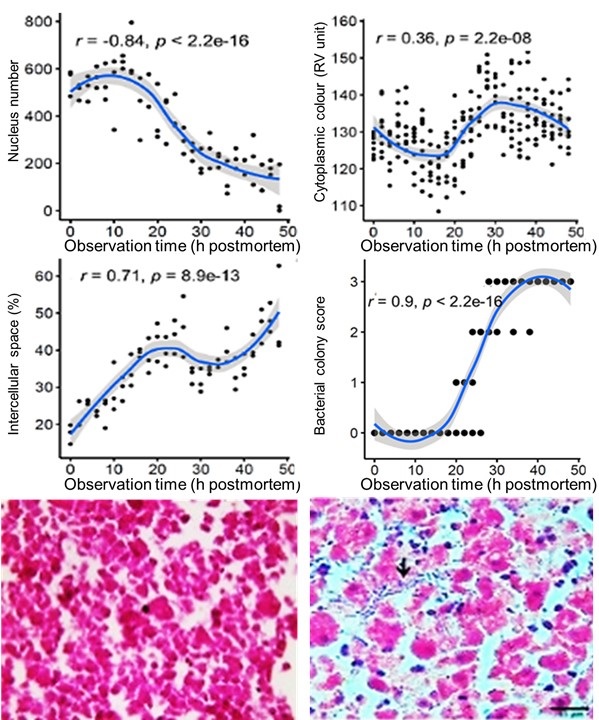
Veterinary forensic science has not received adequate attention in Indonesia; therefore, its development needs to increase. This research aimed to study veterinary forensic science through histopathology of broiler chicken livers 48 h after death. Seventy-five broiler chickens aged 7 days were euthanized and divided into 25 groups based on post-euthanasia necropsy (n=3). Chicken cadavers were necropsied every 2 h postmortem, and livers were collected to prepare histopathological sections and stained with haematoxylin-eosin (HE). Liver histopathology evaluation and the results obtained were analysed using ImageJ software version 1.53a. The relationship between histopathological variables and postmortem time was analysed using the Pearson’s method. The results showed that decay began at the 18th h postmortem which was marked by the number of hepatocyte cell nuclei. At the 20th hour, putrefactive bacteria were found, and at the 22nd hour, there was an increase in the distance between the hepatocytes. Based on the Pearson correlation value, the number of hepatocyte cell nuclei, the distance between hepatocytes, and the presence of putrefactive bacteria have a strong to very strong relationship with postmortem time; therefore, these results can be used to determine the time of death (postmortem interval)
Downloads
References
Brooks JW. 2018. Veterinary Forensic Pathology. Pennsylvania (US): Springer. https://doi.org/10.1007/978-3-319-67175-8
Byard RW, Boardman W. 2011. The potential role of forensic pathologists in veterinary forensic medicine. Forensic Science, Medicine, and Pathology. 7:231-232. https://doi.org/10.1007/s12024-011-9241-x | PMid:21499938
Dettmeyer RB. 2018. Forensic Histopathology: Fundamental and Perspectives. USA: Springer. https://doi.org/10.1007/978-3-319-77997-3 | PMCid:PMC5818275
DiMaio VJ, DiMaio DJ. 2001. Forensic pathology. Florida: CRC Press.
Guo J, Fu X, Liao H, Hu Z, Long L, Yan W, Zha L. 2016. Poten-tial use of bacterial community succession for estimating post-mortem interval as revealed by high through put sequencing. Scientific Reports. 6:24197. https://doi.org/10.1038/srep24197 | PMid:27052375 PMCid:PMC4823735
Huang X, Xiong G, Chen X, Liu R, Li M, Ji L, Zhang X. 2021. Autolysis in crustacean tissues after death: A case study using the Procambarus clarkii hepatopancreas. BioMed Research International. 2021:1-8. https://doi.org/10.1155/2021/2345878 | PMid:33521126 PMCid:PMC7817300
MacKenzie JM, Path FRC. 2014. Examining the decomposed brain. The American Journal of Forensic Medicine and Pathology. 35(4):265-270. https://doi.org/10.1097/PAF.0000000000000111 | PMid:25384305
Moraleda V, Gómez-Catasús J, Schuster C, Carrascal LM. 2022. Decomposition stages as a clue for estimating the post-mortem interval in carcasses and providing accurate bird collision rates. Scientific Reports. 12(1):16188. https://doi.org/10.1038/s41598-022-20628-3 | PMid:36171410 PMCid:PMC9519910
Paczkowski S, Schütz S. 2011. Post-mortem volatiles of vertebrate tissue. Applied Microbiology and Biotechnology. 91(4):917-935. https://doi.org/10.1007/s00253-011-3417-x | PMid:21720824 PMCid:PMC3145088
Copyright (c) 2023 CC-BY-SA

This work is licensed under a Creative Commons Attribution-ShareAlike 4.0 International License.
Authors who publish with this journal agree to the following terms:
1. Authors retain copyright and grant the journal right of first publication with the work simultaneously licensed under a Creative Commons Attribution License that allows others to share the work with an acknowledgement of the work's authorship and initial publication in this journal.
2. Authors are able to enter into separate, additional contractual arrangements for the non-exclusive distribution of the journal's published version of the work (e.g., post it to an institutional repository or publish it in a book), with an acknowledgement of its initial publication in this journal.
3. Authors are permitted and encouraged to post their work online (e.g., in institutional repositories or on their website) prior to and during the submission process, as it can lead to productive exchanges, as well as earlier and greater citation of published work (See The Effect of Open Access).
.jpg)